Breast lump investigation

Published by Bupa's Health Information Team, March 2010.
This factsheet is for people who are having a breast lump investigated, or who would like information about it.
Breast lump investigation is any technique used to diagnose breast conditions, including imaging and biopsy procedures.
You will meet the doctor carrying out your procedure to discuss your care. It may differ from what is described here as it will be designed to meet your individual needs.
About breast lump investigation
Preparing for your triple assessment
What happens during triple assessment
What to expect afterwards
Recovering from triple assessment
What are the risks?
About breast lump investigation
A process called triple assessment is used to diagnose breast lumps. There are three stages in triple assessment.
- Examination – a doctor or nurse asks about your medical history and examines your breast.
- Imaging – pictures of the inside of your breast are created using ultrasound or X-rays.
- Biopsy – a sample of your breast tissue is removed and sent to a laboratory for testing to determine whether the cells are cancerous (malignant) or not cancerous (benign).
The results of the triple assessment can help your doctor decide if you need any further treatment.
The procedures described here are usually done in an out-patient breast clinic at a hospital.
Preparing for triple assessment
Your doctor will discuss with you what will happen before, during and after your triple assessment, and any pain you might have. This is your opportunity to understand what will happen, and you can help yourself by preparing questions to ask about the risks, benefits and any alternatives to the procedures. This will help you to be informed, so you can give your consent for the procedures to go ahead, which you may be asked to do by signing a consent form.
What happens during triple assessment
Breast examination
You will need to remove all your clothes above your waist. Your doctor will examine your breasts and armpits and press gently on your skin to feel for any changes in texture.
Breast imaging
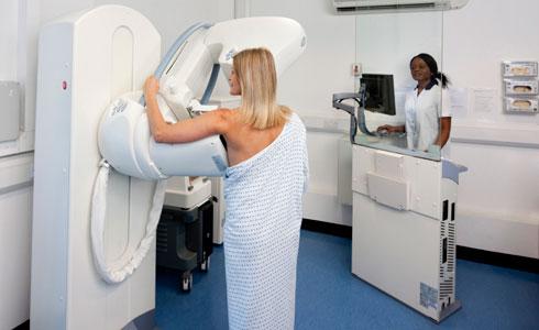
In order to see where the lump is, you may need to have a picture taken of the inside of your breast. This is called imaging. Imaging is usually done in the X-ray department by a radiologist (a doctor who specialises in using imaging methods to diagnose medical conditions) or a radiographer (a health professional trained to perform imaging procedures). There are two imaging procedures commonly used.
- A mammography uses X-rays to create a picture of your breast. Mammography is usually done while you’re standing up. Your breast will be pressed between two plastic plates to keep it still. Some women find the pressure of these plates uncomfortable.
- An ultrasound uses sound waves to produce an image of the inside of your breast. Your radiologist or radiographer will put gel on your breast and then move a sensor over your skin. You will probably be asked to sit or lie on an examination couch for the scan.
Breast biopsy
A breast biopsy is a small sample of tissue that is taken from your breast. The sample will be sent to a laboratory for testing to determine the type of cells and if these are cancerous or not.
A breast biopsy may be done under local anaesthesia. This completely blocks pain from your breast and you will stay awake during the procedure. The local anaesthetic is injected into your breast. The injection may sting briefly.
There are several different biopsy procedures including fine needle aspiration, core biopsy, vacuum assisted core biopsy and open biopsy. Your doctor will explain which procedure is most suitable for you.
Fine needle aspiration
Your doctor will collect cell samples from your breast using a fine needle. He or she will pass the needle through the skin of your breast, into the lump or breast tissue being examined and draw cells out into a syringe. Sometimes ultrasound or X-rays are used to help guide your doctor to the area that needs to be checked.
Core biopsy
Your doctor will collect breast tissue samples using a hollow needle. Your doctor will pass the needle through your breast to the area to be checked. Sometimes ultrasound or X-rays are used to help your doctor guide the needle. He or she will then release a spring in the needle and breast tissue will be collected inside the hollow cylinder. The needle may need to be inserted several times to get more than one sample of breast tissue. The spring action is quite sudden and can surprise you the first time.
Vacuum assisted core biopsy (VACB)
Your doctor will collect breast tissue samples using a special, hollow probe attached to a gentle vacuum pump. Your doctor will make a small cut in your breast over the area being examined and insert the probe. The probe will suck some of your breast tissue into a cylinder. More than one sample can be taken without your doctor removing the probe.
VACB is useful for removing larger samples of breast tissue and sometimes a whole lump can be removed in this way. Ultrasound may be used to make sure the correct breast tissue is removed.
Open biopsy
You will have a minor operation to remove the whole lump under a general anaesthetic. This means you will be asleep during the procedure. Most hospitals will do the biopsy as a day case but you may need to stay a night in hospital. An open may also be referred to as an excision biopsy.
What to expect afterwards
You will be able to go home when you feel ready. Your nurse will give you advice about caring for your breasts before you go home.
If you have any pain, take over-the-counter painkillers such as paracetamol or ibuprofen. Always read the patient information that comes with your medicine and if you have any questions, ask your pharmacist for advice.
It may be possible for your doctor to diagnose your breast lump on the same day. However, some tests can take a few days to carry out. Your clinic will get your results to you as soon as possible – ask your doctor or breast care nurse when to expect your results. Often, you will be invited to a follow-up appointment with your doctor and breast care nurse to discuss your biopsy results.
Recovering from triple assessment
After a triple assessment you should be able to return to your usual activities straight away, but don't do any strenuous exercise or lifting for the first 24 hours.
If you have any pain that can't be controlled with over-the-counter painkillers, a high temperature or your breast feels unusually hot to touch, contact the hospital as you may have developed an infection.
What are the risks?
A triple assessment is commonly performed and generally safe. However, in order to make an informed decision and give your consent, you need to be aware of the possible side-effects and the risk of complications.
Side-effects
Side-effects are the unwanted but mostly temporary effects you may get after having the procedures.
Your breast may feel sore and bruised for a few days, depending on the type of biopsy you have. It's unusual to have any noticeable scars after a breast biopsy, but on rare occasions you may develop a small scar. This depends on the size and type of biopsy you have.
Complications
Complications are when problems occur during or after the procedures.
Occasionally, some people faint if they are having a biopsy done standing up. If this happens, the team at the hospital will look after you until you feel ready to go home.
Your doctor will be very experienced at taking breast biopsies but, even so, the biopsy may not be successful. If this happens you may need to have another biopsy or an operation to remove the abnormal breast tissue or lump.
The exact risks are specific to you and differ for every person, so we haven't included statistics here. Ask your doctor to explain how these risks apply to you.
This section contains answers to common questions about this topic. Questions have been suggested by health professionals, website feedback and requests via email.
Does a breast lump mean I have breast cancer?
What causes nipple discharge?
Will the breast biopsy hurt?
Does a breast lump mean I have breast cancer?
Finding a lump in your breast doesn’t always mean you have cancer. Most breast lumps aren’t cancerous and may, instead, be a cyst or fibroadenoma.
Explanation
Breast cysts are sacs of fluid that build up in your breast tissue. Fluid can build up in your breast tissue as a result of hormonal changes. For example, it's normal to have lumpy, tender breasts just before your period, especially near your armpits. Cysts are most common in women who are over 35, before they go through the menopause. Breast cysts can be painful and usually move easily under your skin. If a cyst is causing you pain or discomfort the fluid can be drained using a needle.
Fibroadenomas are harmless overgrowths of your breast tissue. They are usually diagnosed after a needle biopsy. Fibroadenomas are most common in women under 30 but can occur at any age. They are often painless and move easily under your skin. Most fibroadenomas will slowly shrink in size and don’t require treatment, but if the lump is getting bigger you can have it removed.
Check your breasts a week after your period because this is when they are softest. If you find a lump, it's best to have it checked by your GP. If your doctor has any concerns, he or she will refer you to a specialist.
It’s also important for men to check their breasts regularly. If you see any change or feel a lump, it's best to have it checked by your GP.
Further information
- Breast Cancer Care
0808 800 6000
www.breastcancercare.org.uk
- Breakthrough Breast Cancer
08080 100 200
www.breakthrough.org.uk
Sources
- Referral to a breast clinic. Breastcancer Care. www.breastcancercare.org.uk, accessed 7 December 2009
- Are all lumps cancer? Breakthrough Breast Cancer. www.breakthrough.org.uk, accessed 7 December 2009
- Fibroadenoma of breast. GP Notebook. www.gpnotebook.co.uk, accessed 7 December 2009
- Fibroadenoma: management. GP Notebook. www.gpnotebook.co.uk, accessed 7 December 2009
- Breast cysts. Breastcancer Care. www.breastcancercare.org.uk, accessed 7 December 2009
What causes nipple discharge?
Nipple discharge is usually caused by swelling of the ducts underneath your nipple.
Explanation
Nipple discharge is fluid that comes from your nipples. All women have fluid in their breasts and it's possible that some of this may come out, particularly during vigorous exercise or sexual activity. Most often the discharge comes from the milk ducts in your breast. It may range in colour from white or pale yellow to green or blue/black and may come from one or both of your nipples.
You may get a blood-stained discharge if you have a duct infection or if you develop a wart in the ducts, called papilloma.
As you grow older, the ducts widen and sometimes they can get blocked, causing a thick yellow or blood-stained nipple discharge. This is more common in women reaching the menopause. A skin infection on the surface of your nipple can also cause your nipple to leak fluid.
If you notice any blood-stained or unusual discharge coming from your nipples, it's best to have it checked by your GP.
Further information
- Breakthrough Breast Cancer
08080 100 200
www.breakthrough.org.uk
Sources
- Duct ectasia. Breastcancer Care. www.breastcancercare.org.uk, accessed 7 December 2009
- Intraductal papilloma. Breastcancer Care. www.breastcancercare.org.uk, accessed 7 December 2009
Will the breast biopsy hurt?
How much discomfort you feel during a biopsy depends on the type of breast biopsy you have and whether your doctor gives you an injection of local anaesthetic before the procedure. Neither a fine needle biopsy nor a core biopsy should be painful if you have a local anaesthetic beforehand.
Explanation
A fine needle biopsy involves only a small sample of tissue being taken from your breast. You may be given an injection of local anaesthetic before the procedure to block pain from the area. Ask your doctor whether or not you will be given an anaesthetic. A fine needle is passed through the skin into the breast area being examined (usually just once) and cells are drawn out into a syringe attached to the needle. If you haven’t had a local anaesthetic, you may feel a slight sting similar to having an injection.
A core biopsy involves removing a larger sample of your breast tissue and it can be painful. Your doctor will usually give you an injection of local anaesthetic before the biopsy to help minimise any discomfort. The anaesthetic is injected into your breast and this may sting briefly. The anaesthetic completely blocks pain from your breast and you will stay awake during the procedure. You may need pain relief after the effects of the local anaesthetic wear off.
Further information
- Breakthrough Breast Cancer
08080 100 200
www.breakthrough.org.uk
Sources
- Visiting a breast clinic. Breakthrough Breast Cancer. www.breakthrough.org.uk, accessed 7 December 2009
- Breast cancer evaluation. Emedicine. www.emedicine.medscape.com, accessed 7 December 2009
- Breast clinic investigations. Breakthrough Breast Cancer. www.breastcancercare.org.uk, accessed 7 December 2009
- Breast cancer tests. Cancer Research UK. www.cancerhelp.org.uk, accessed 7 December 2009
- Fine needle aspiration cytology (breast). GP Notebook. www.gpnotebook.co.uk, accessed 7 December 2009
Related topics
Benign breast lumps
Breast awareness
Breast cancer
Breast lump removal
Menopause
This information was published by Bupa’s Health Information Team and is based on reputable sources of medical evidence. It has been peer reviewed by Bupa doctors. The content is intended for general information only and does not replace the need for personal advice from a qualified health professional.
Publication date: March 2010.
Breast lump investigation factsheet
Please visit the breast lump investigation health factsheet for more information.
Related topics
Benign breast lumps
Breast awareness
Breast cancer
Breast lump removal
Local anaesthesia and sedation
Mammography if you have breast symptoms
Further information
- Macmillan cancer support
0808 808 0000
www.macmillan.org.uk
- Breast Cancer Care
0808 800 6000
www.breastcancercare.org.uk
Sources
- Visiting a breast clinic. Breakthrough Breast Cancer. www.breakthrough.org.uk, accessed 7 December 2009
- Breast cancer evaluation. Emedicine. www.emedicine.medscape.com, accessed 7 December 2009
- Early and locally advanced breast cancer: diagnosis and treatment. National Institute for Health and Clinical Excellence (NICE), February 2009. www.nice.org.uk
- How breast cancer is diagnosed. Macmillan Cancer Support. www.macmillan.org.uk, accessed 7 December 2009
- Breast clinic investigations. Breakthrough Breast Cancer. www.breastcancercare.org.uk, accessed 7 December 2009
- Breast cancer - suspected. Clinical Knowledge Summaries. www.cks.nhs.uk, accessed 7 December 2009
- Breast cancer tests. Cancer Research UK. www.cancerhelp.org.uk, accessed 7 December 2009
- Berg WA, Blume JD, Cormack JB et al. Combined screening with ultrasound and mammography vs mammography alone in women at elevated risk of breast cancer. JAMA 2008; 299(18):2151-63. doi: 10.1001/jama.299.18.2151
- Fine needle aspiration cytology (breast). GP Notebook. www.gpnotebook.co.uk, accessed 7 December 2009
- Open biopsy (breast). GP Notebook. www.gpnotebook.co.uk, accessed 7 December 2009
- Breast calcifications. Breastcancer Care. www.breastcancercare.org.uk, accessed 7 December 2009
- James JJ, Wilson AR, Evans AJ et al. The use of a short-acting benzodiazepine to reduce the risk of syncopal episodes during upright stereotactic breast biopsy. Clin Radiol 2005; 60(3):394-6. doi:10.1016/j.crad.2004.09.003
This information was published by Bupa’s Health Information Team and is based on reputable sources of medical evidence. It has been peer reviewed by Bupa doctors. The content is intended for general information only and does not replace the need for personal advice from a qualified health professional.
Publication date: March 2010.


