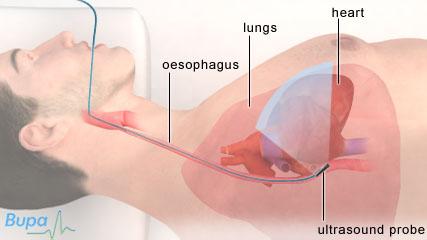
Transoesophageal echocardiogram

An echocardiogram uses ultrasound to produce moving real-time images of the inside of your heart. In a TOE, the ultrasound probe is placed in your oesophagus (the pipe that goes from the mouth to the stomach), to take the images of your heart. It can help to diagnose heart problems.
About transoesophageal echocardiogram
What are the alternatives?
Preparing for a transoesophageal echocardiogram
What happens during a transoesophageal echocardiogram
What to expect afterwards
What are the risks?
About transoesophageal echocardiogram
An echocardiogram is used to check the structure of your heart and how well it’s working. It uses an ultrasound probe to get a moving picture of your heart.
Transoesophageal means that the images are taken from inside your oesophagus, the pipe that joins your mouth to your stomach. An echocardiogram done this way gives a clearer view of your heart than a standard echocardiogram, which takes the images through your chest. The test can also give a detailed view of the heart valves. You may have a TOE to check for:
• problems with your heart valves
• infection of the lining inside your heart
• swelling of, or tears in, your aorta (the largest artery in your body)
• damage to your heart muscle
• blood clots
A transoesophageal echocardiogram can also be used to take images of your heart during heart procedures, such as surgery and cardiac catheterisation.
What are the alternatives?
There are a number of alternatives to having a transoesophageal echocardiogram. Some of these are listed below.
• Electrocardiogram (ECG). This is a test that measures the electrical activity of your heart to see how well it’s working.
• A standard echocardiogram. For this type of echocardiogram, the ultrasound probe is moved over your chest.
• A stress echocardiogram. This is similar to a standard echocardiogram, but it’s carried out when your heart is under stress (either after exercise or after being given a medicine)
• Radionuclide test. During this test you’re injected with a harmless, radioactive substance when you’re resting and when your heart is under stress. As it travels through your heart, the radioactive substance is seen with a special camera.
• Cardiac MRI (magnetic resonance imaging) test. An MRI scan uses magnets and radiowaves to produce images of the inside of the body.
Preparing for a transoesophageal echocardiogram
A TOE is usually carried out in hospital by a cardiologist (a doctor specialising in conditions of the heart and blood vessels). You will be asked to attend the hospital as an out-patient and should follow any instructions given to you in your appointment letter.
You shouldn’t eat or drink anything for at least six hours before the test. If you have diabetes, you may be asked to not take your diabetes medication before the test. You should take any other medication as usual. You will also need to arrange for someone to take you home after the test.
Your doctor will discuss with you what will happen before, during and after your procedure, and any pain you might have. This is your opportunity to understand what will happen, and you can help yourself by preparing questions to ask about the risks, benefits and any alternatives to the procedure. This will help you to be informed, so you can give your consent for the procedure to go ahead, which you may be asked to do by signing a consent form.
What happens during a transoesophageal echocardiogram
You may have to stay in hospital for a few hours for a TOE. The procedure takes place in a private room, and you may be asked to change into a hospital gown. You will have a local anaesthetic sprayed on the back of your throat to numb it and to help you to swallow the ultrasound probe. You may also have a sedative injection, which will help you to relax and relieve any anxiety. You will be given a plastic mouth guard to protect your teeth.
You will be asked to lie on your side and to swallow a small probe, which is on the end of a flexible tube. You will be able to breathe normally. The probe gives out ultrasound waves, which bounce off the structures of your heart and are picked up by the echocardiogram machine. The images of your heart will be displayed on a screen. When the test is finished the doctor will gently take the tube out.
Sometimes a contrast liquid may be injected into your vein during the test. This helps to show certain parts of your heart more clearly.
The procedure itself usually takes 15 to 20 minutes. You will normally be given some time to rest after it has finished.
What to expect afterwards
If you have had a sedative, you may need to stay in hospital for a couple of hours. Sedation temporarily affects your co-ordination and reasoning skills, so you must not drive, drink alcohol, operate machinery or sign legal documents for 24 hours afterwards. If you’re in any doubt about driving, contact your motor insurer so that you’re aware of their recommendations, and always follow your doctor’s advice.
You shouldn’t eat or drink anything for the first few hours after a TOE, until your local anaesthetic has worn off.
The results of your echocardiogram are usually sent to your cardiologist or to the doctor who requested the test, for example your GP. He or she will discuss the results with you at your next appointment.
What are the risks?
Transoesophageal echocardiogram is commonly performed and generally safe. However, in order to make an informed decision and give your consent, you need to be aware of the possible side-effects and the risk of complications of this procedure.
Side-effects
Side-effects are the unwanted but mostly temporary effects you may get after having the procedure.
The back of your throat may feel sore and swallowing may be uncomfortable after a TOE. This should wear off very quickly.
Complications
This is when problems occur during or after the procedure. Most people aren’t affected.
You may have small injuries to your mouth or oesophagus. These don’t normally need any treatment.
There is a small risk of damage to your teeth. If you have dental crowns or bridges, there is also a small chance that these may get damaged. You should tell your doctor if you have a dental plate (false teeth) and remove it before the investigation.
If you had a contrast liquid injected during the test, there is a small risk of bruising around the place when the injection was given. There is also a very small risk of an allergic reaction to the contrast liquid.
The exact risks are specific to you and differ for every person, so we haven't included statistics here. Ask your doctor to explain how these risks apply to you.
Answers to questions about transoesophageal echocardiogram
This section contains answers to common questions about this topic. Questions have been suggested by health professionals, website feedback and requests via email.
What if I can’t swallow the probe?
Why am I having a transoesophageal echocardiogram and not a normal echocardiogram?
How will my doctor know if there is a problem with my heart?
What will happen after I get the results of my transoesophageal echocardiogram?
What if I can’t swallow the probe?
A local anaesthetic is sprayed onto the back of your throat before the echocardiogram begins, which helps to make it easier for you to swallow the probe. You may also have a sedative to help you relax. This means it's very unlikely that you won't be able to swallow the probe.
Explanation
Some people do find it difficult to swallow the probe. However, a local anaesthetic spray helps to numb the back of your throat. If you do find swallowing the probe difficult, your doctor can give you a sedative medicine. This relieves anxiety and helps you relax.
You should tell your doctor before the test if you have had any surgery to your throat or neck, problems swallowing or if you have ever coughed up blood. Your doctor may suggest that you have an alternative test.
Why am I having a transoesophageal echocardiogram and not a normal echocardiogram?
A transoesophageal echocardiogram produces more detailed pictures of your heart than a normal echocardiogram. This makes it quicker and easier for your doctor to diagnose any heart problems you may have. It’s also much better for investigating artificial heart valves and looking for a blood clot (thrombus) in your heart.
Explanation
A routine echocardiogram is done with an ultrasound probe moved over the front of your chest. This is sometimes called a transthoracic view. Although it produces good pictures of your heart, the sound waves have to pass through your skin, fat, bone, tissue and air of your ribcage and lungs. This means that the pictures aren’t as clear as in a transoesophageal echocardiogram (TOE), where the probe is placed very close to the back of the heart.
How will my doctor know if there is a problem with my heart?
A transoesophageal echocardiogram (TOE) is usually able to produce very detailed pictures of the structures inside your heart, which allows your doctor to identify any problems.
Explanation
If your doctor is checking for problems with your heart valves, he or she will look at the shape of the valves, how they are moving and whether they are calcified (have a build-up of calcium deposits). By measuring how fast your blood is flowing, your doctor will be able to see whether the valves have become narrow or are leaking.
If you have an infection of your heart valves, the TOE may show this. If you have damage to your heart muscle, the lower chambers of your heart (the ventricles) may appear thinned, thickened, scarred or enlarged.
What will happen after I get the results of my transoesophageal echocardiogram?
Your doctor will discuss your treatment options with you, based on the results of your transoesophageal echocardiogram (TOE) together with any other tests you have had. Depending on the results, you may need to have treatment such as medicines or surgery.
Explanation
An echocardiogram is just one test that doctors use to assess how well your heart is working. You may have other tests including an electrocardiogram (ECG), a chest X-ray and blood and urine tests.
Your doctor may diagnose a problem with your heart using the results of all these tests. However, your TOE may also rule out a problem with your heart, or you may need further tests before a diagnosis can be made.
If tests do show up a problem with your heart, your doctor will discuss your treatment options with you. Depending on the problem identified, you may be prescribed medicine for your condition or be advised to have surgery.
Keywords Transoesophageal echocardiogram, TOE, echocardiogram, oesophagus
Further information
• British Cardiac Patients Association
01223 846845
www.bcpa.co.uk
• British Heart Foundation
0300 330 3311
www.bhf.org.uk
Sources
• HIS9 Tests for heart conditions. British Heart Foundation, published March 2009..
• Transesophageal echocardiography. American Heart Association. www.americanheart.org, accessed 9 March 2010
• Echocardiogram. British Cardiac Patients Association. www.bcpa.co.uk, published 2008
• Echocardiogram. British Heart Foundation. www.bhf.org.uk, published July 2009
• Transoesophageal echocardiogram (animation). British Heart Foundation. www.bhf.org.uk, accessed 9 March 2010
• Transoesophageal echocardiogram. British Cardiovascular Society. www.bcs.com, accessed 8 March 2010
Related topics
• Diagnosing heart conditions
• Echocardiogram
• Electrocardiogram
• Heart attack
• Heart failure
• Heart valve disease
• Transoesophageal echocardiogram


