Ultrasound in pregnancy
This factsheet is for women who are having an ultrasound scan during pregnancy, or who would like information about it.
An ultrasound uses sound waves to produce an image of the inside of your body. It's used during pregnancy to monitor your baby's growth and check for physical abnormalities.
You will meet the obstetrician or sonographer carrying out your procedure to discuss your care. It may differ from what is described here as it will be designed to meet your individual needs. Details of the procedure may also vary from country to country.
About ultrasound in pregnancy
Preparing for an ultrasound scan
What happens during an ultrasound scan
What to expect afterwards
What are the risks?
About ultrasound in pregnancy
An ultrasound uses high-frequency sound waves and their echoes to create moving three-dimensional (3D) or four-dimensional (4D) images of your growing baby. The pictures (scans) are black, white and grey and will be displayed on a monitor for you to see during the procedure.
Ultrasound scans in pregnancy are usually performed by a sonographer or an obstetrician. An obstetrician is a doctor who specialises in pregnancy and childbirth. A sonographer is a technician who is specially trained to take ultrasound scans.
There are different reasons for doing ultrasound scans at different stages during your pregnancy. When you're pregnant, you're likely to be offered at least two scans – a 'dating scan' to primarily check your due date, and a 'fetal anomaly scan' to check that your baby is developing normally. You may be offered additional scans if you're at a higher risk of medical problems or have a family history of certain medical conditions that may affect your pregnancy.
Dating scan
You will normally be offered an ultrasound scan between eight and 14 weeks of your pregnancy to check when your baby is due. This can help you to monitor important milestones during your pregnancy. This scan will also tell you if you're expecting more than one baby. If you have the scan between 11 and 13 weeks of getting pregnant, your baby can also be screened for Down's syndrome.
Fetal anomaly scan
You will usually be offered another scan to check your baby's development between 18 and 20 weeks of your pregnancy. During this scan, your obstetrician or sonographer will check for abnormalities. He or she will check your baby's heart, brain, kidneys, liver and spine, and measure the size of their arms, legs and head.
Your sonographer will also check the position of the placenta, which provides vital nutrients and oxygen-rich blood to your baby. If the placenta is lying unusually low in your womb, this is called a marginal, or low-lying placenta. This will usually resolve before your baby is born, but if it doesn't, it is called placenta praevia and you may need to have a caesarean delivery – an operation to deliver your baby through your abdomen (tummy).
Other ultrasound scans in pregnancy
Ultrasound is used in other procedures that you may be offered during your pregnancy. For example, your obstetrician or sonographer may use an ultrasound to guide a fine needle through your abdomen to collect a sample of amniotic fluid that surrounds your baby for an amniocentesis test or to collect tissue samples from your placenta for chorionic villus sampling.
You may have other scans during pregnancy if your routine scans or antenatal appointments suggest there may be a problem with your baby or the placenta. For example, you might have more ultrasound scans if:
- your fetal anomaly scan showed you have a low-lying placenta
- you have diabetes and are at risk of having a large-for-gestational-age baby
- your midwife thinks your baby may be in a breech position (bottom-down rather than head-down)
- you have vaginal bleeding
Doppler ultrasound
A Doppler ultrasound monitors flow in your blood vessels and is sometimes used to check how well your placenta is functioning as this can affect your baby's growth and development. This isn't a routine test – you will only be offered it if your obstetrician thinks there might be a problem with the placenta.
Fetal echocardiogram
Fetal echocardiogram is a type of Doppler ultrasound to examine your baby's heart before birth. It's usually done at around 18 to 23 weeks by scanning through your abdomen. You will only be offered a fetal echocardiogram if a routine scan shows abnormalities, or if your baby is at risk of having heart problems, such as congenital heart disease.
Preparing for an ultrasound scan
A doctor or midwife will arrange your ultrasound scans. You usually have the scan in an outpatient department in hospital or in a clinic.
Your obstetrician or sonographer will explain how to prepare for your procedure. In early pregnancy you may need to have a full bladder, so you will be asked to drink fluids about an hour before the scan. A full bladder will help to lift your large bowel out of your pelvis so that your womb (uterus) can be seen more easily.
Your obstetrician or sonographer will discuss with you what will happen before, during and after your procedure, and any discomfort you might have. This is your opportunity to understand what will happen, and you can help yourself by preparing questions to ask about the risks, benefits and any alternatives to the procedure. This will help you to be informed, so you can give your consent for the procedure to go ahead, which you may be asked to do by signing a consent form.
What happens during an ultrasound scan
An ultrasound scan usually takes 10 to 15 minutes to perform. A Doppler scan or fetal echocardiogram may take longer depending on the investigation.
The ultrasound scanner looks a bit like a home computer system. There is a hard drive, keyboard and a display screen. There is a sensor that your obstetrician or sonographer will hold and this will send out sound waves and pick up the returning echoes. Pictures of your baby will be displayed on a monitor – these are constantly updated so the scan can show your baby's movements.
You may have the ultrasound scan through your vagina or abdomen depending on how many weeks pregnant you are. Both the dating scan and the fetal anomaly scan are usually abdominal scans.
Vaginal scan
This method is used if the scan is being done in early pregnancy (before about 12 weeks) when the embryo is very small. A vaginal scan gives a better view compared to an abdominal scan at this stage.
You will be asked to lie on your back and your obstetrician or sonographer will gently insert a lubricated sensor (the size of a tampon) into your vagina. The sensor will usually be covered with a condom. Please tell your examiner if you suffer from a latex allergy, so that a suitable condom is used.
Abdominal scan
This method is usually used for scans after about 10 weeks of pregnancy.
You will be asked to lie down on your back. Your sonographer or obstetrician will rub clear gel onto your skin on your lower abdomen. The gel allows the sensor to slide easily over your skin and helps to produce clearer pictures. Your obstetrician or sonographer will hold the sensor firmly against your skin and will move it over the surface. If you look at the screen, you will see a picture of your baby.
You can go home when the scan is finished. Permanent copies of your scan will be stored on a computer, a disc or printed. Your obstetrician or sonographer may give you a printed copy of the scan to take home with you after having routine scans. Some clinics and hospitals save the scans on a DVD for you to take home – ask your obstetrician or sonographer for more information.
What to expect afterwards
Your sonographer or obstetrician may explain the details of your ultrasound scan to you during or straight after your scan. Sometimes, the results of your scan will be sent to your midwife or doctor who requested it and you will need to make an appointment to find out the results.
You will usually be able to go home when you feel ready.
What are the risks?
An ultrasound examination is painless and safe. It doesn't use radiation or have any harmful effects that may harm your baby. It's considered safe to use during pregnancy.
This section contains answers to common questions about this topic. Questions have been suggested by health professionals, website feedback and requests via email.
What happens if my ultrasound scan shows an ectopic pregnancy?
How soon can an ultrasound scan determine whether my baby is a boy or girl?
What is the purpose of my 12-week ultrasound pregnancy scan?
What happens if my ultrasound scan shows an ectopic pregnancy?
Answer
An ectopic pregnancy must be treated because it's a life-threatening condition and the embryo will not survive.
Explanation
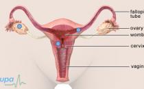
An ultrasound scan can show an ectopic pregnancy as early as five weeks. An ectopic pregnancy is when the embryo attaches outside the womb, usually to the fallopian tube and sometimes to the ovary or cervix.
An embryo that attaches outside the womb can’t develop normally and can damage the organ it's attached to, causing severe bleeding and putting the woman’s life at risk. This is why a confirmed ectopic pregnancy needs to be ended, using either medicines or surgery.
Most ectopic pregnancies are treated using a medicine called methotrexate. This stops new cells from being produced and so stops the growth of the pregnancy. Methotrexate is usually given as an injection.
The embryo can be surgically removed using keyhole or open surgery. In keyhole surgery, special instruments are passed into the abdomen through small cuts. These instruments are used to examine and remove the ectopic pregnancy. For open surgery, the surgeon makes a single cut into the abdomen and removes the embryo. Treating ectopic pregnancy by surgery is usually a medical emergency.
Further information
-
The Ectopic Pregnancy Trust
020 7733 2653
www.ectopic.org.uk
Sources
- Ectopic pregnancy. Emedicine. www.emedicine.medscape.com, accessed 30 September 2009
- Ectopic pregnancy: treatment. www.emedicine.medscape.com, accessed 30 September 2009
How soon can an ultrasound scan determine whether my baby is a boy or girl?
Answer
An ultrasound scan can check the sex of your baby from around 15 weeks of pregnancy.
Explanation
Ultrasound can check the sex of your baby from around four months of pregnancy. However, it is not completely accurate because it depends on the position of your baby and the skill of your sonographer. You may be able to find out at your routine anomaly scan, but some hospitals will not tell you the sex of your baby unless it is for medical reasons.
If you need to know the sex of your baby for medical reasons, you may be offered amniocentesis or chorionic villus sampling (CVS). These tests can help determine the sex of your baby and check for a range of genetic disorders.
Amniocentesis involves taking a sample of amniotic fluid that surrounds your baby in the womb. Amniocentesis has a small risk of causing a miscarriage. This is why it is usually offered only to women when screening tests show they have a higher risk of having a baby with a genetic disorder, or women over 35 years old as the chance of having a Down's syndrome baby increases with the mother’s age.
Chorionic villus sampling (CVS) involves removing tiny tissue samples from the placenta. CVS is usually done at 10 to 13 weeks of pregnancy. The procedure has a slightly higher risk of miscarriage compared to amniocentesis. It is also not as accurate as amniocentesis.
Further information
-
The Royal College of Obstetricians and Gynaecologists
020 7772 6200
www.rcog.org.uk
Sources
- Key facts: what is haemophilia? Haemophilia Society. www.haemophilia.org.uk, accessed 30 September 2009
- Amniocentesis and chorionic villus sampling. Royal College of Obstetricians and Gynaecologists, 2005. www.rcog.org.uk
What is the purpose of my 12-week ultrasound pregnancy scan?
Answer
You will usually be offered an ultrasound scan at about 10 to 14 weeks of pregnancy. It's often called a dating scan because it's done to check how many weeks pregnant you are and estimate your expected due date. During this scan, your baby can be screened for Down's syndrome.
Explanation
Your midwife or doctor can tell how far into your pregnancy you are by measuring your baby's length from top of head to rump. This is called the crown-rump length (CRL). On average your baby is about 3–8cm long at 10 to 14 weeks of pregnancy. Your baby's face is well formed and his or her eyelids are closed. Your baby can open and close his or her mouth and frown. The arms and legs are long and thin and the baby can make a fist and curl toes. Your baby's sex organs are developed but they are too small to see on the scan.
The amount of fluid in a fold behind your baby's neck can be measured to assess the risk of Down's syndrome. This is called the nuchal translucency test. The more fluid is present, the greater the chance the baby has Down’s syndrome. People with Down's syndrome have an extra chromosome 21. They have characteristic physical and mental features such as learning difficulties, particular facial features and heart problems.
If the test indicates a higher risk, you will be offered tests such as amniocentesis or chorionic villus sampling (CVS), which can tell you if your baby has Down’s syndrome.
Further information
-
Down's Syndrome Association
0845 230 0372
www.downs-syndrome.org.uk -
The Royal College of Obstetricians and Gynaecologists
020 7772 6200
www.rcog.org.uk
Sources
- Antenatal care: routine care for the healthy pregnant woman. National Institute for Health and Clinical Excellence (NICE), 2008. www.nice.org.uk
- Fetal development. National Library of Medicine. www.nlm.nih.gov, accessed 30 September 2009
- Your second trimester: Nuchal translucency screening. Emma’s Diary, Royal College of General Practitioners. www.emmasdiary.co.uk, accessed 30 September 2009
Related topics
Ectopic pregnancy
Down’s syndrome
Ultrasound in pregnancy factsheet
Visit the ultrasound in pregnancy health factsheet for more information.
Related topics
- Pelvic and abdominal ultrasound
- Antenatal care
- Stages of pregnancy
Further information
-
National Childbirth Trust (NCT)
0300 330 0772
www.nct.org.uk -
Emma's diary (Royal College of General Practitioners)
www.emmasdiary.co.uk
Sources
- The Pregnancy Book. Department of Health. www.dh.gov.uk, published 2009
- Antenatal care – uncomplicated pregnancy. Prodigy. www.prodigy.clarity.co.uk, published March 2011
- Down's syndrome screening. Map of Medicine. www.mapofmedicine.com, published January 2012
- Antenatal care: routine care for the healthy pregnant woman. National Institute for Health and Clinical Excellence (NICE), June 2010. www.nice.org.uk
- Alfirevic Z, Stampalija T, Gyte GML. Fetal and umbilical doppler ultrasound in normal pregnancy. Cochrane Database of Systematic Reviews 2010, Issue 8. doi: 10.1002/14651858.CD001450.pub3
- Obstetrical ultrasound. RadiologyInfo.org. www.radiologyinfo.org, accessed 8 December 2011
- Antenatal screening for congenital heart disease. British Heart Foundation. www.bhf.org, published March 2009
- Information for patients having an ultrasound scan. Royal College of Radiologists. www.rcr.ac.uk, published December 2010
- What to expect from different types of ultrasound examination. British Medical Ultrasound Society. www.bmus.org, accessed 8 December 2011
- Ectopic pregnancy – suspected. Map of Medicine. www.mapofmedicine.com, published 19 July 2011
- Ectopic pregnancy – management. Map of Medicine. www.mapofmedicine.com, published 19 July 2011
- Ultrasound in pregnancy. The Society of Obstetricians and Gynaecologists of Canada (SOGC). www.sogc.org, published 4 May 2011
- Amniocentesis and chorionic villus sampling. Royal College of Obstetricians and Gynaecologists. www.rcog.org.uk, published June 2010
- Arulkummaran S, Symonds IM, A F. Oxford handbook of obstetrics and gynaecology. Oxford: Oxford University Press; 2004:82–83
Produced by Stephanie Hughes, Bupa Health Information Team, March 2012.


