Electrocardiogram (ECG)
An ECG records the rhythm and the electrical activity of your heart. It's a test used to find out if your heart is healthy.
You will meet the doctor, nurse or technician carrying out your procedure to discuss your care. It may differ from what is described here as it will be designed to meet your individual needs. Details of the procedure may also vary from country to country.
How the heart works
About ECG
Preparing for an ECG
What happens during an ECG
What to expect afterwards
What are the risks?
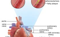
How the heart works
The information on the video provided does not constitute advice on diagnosis or the treatment for heart disease and such advice should always be sought from a doctor or another suitably qualified health professional.
About ECG
An ECG is a simple test to record information about your heartbeat and the rhythm of your heart. An ECG measures the electrical signals that cause your heart to beat. During the test, a number of wires are connected to your arms, legs and chest and these pick up the electrical signals. These signals can be seen on a screen or are traced out on a piece of paper.
You may have an ECG done at a doctor's clinic or in hospital. An ECG is one of the tests you may have if you're taken to hospital as an emergency because you have chest pains or an abnormal heart rate.
There are a number of reasons why you may need to have an ECG. You may have one to check for problems with your heart if you're having symptoms such as dizziness, chest pain or an abnormal heart rate. You may also have one as routine before an operation or part of a health check.
An ECG can show a number of different heart problems including:
• a previous heart attack or a heart attack that is happening at the time of the test
• an enlarged heart that is working under strain
• fast, slow or irregular heartbeats called arrhythmias
There are a number of different types of ECG. These are listed below.
• The standard ECG, which is sometimes called a resting ECG. This is taken while you're resting.
• An exercise ECG, which is taken while you're exercising. This shows how your heart copes under stress. The test can help to find out if you have coronary heart disease (when the arteries to your heart become narrowed). If you have recently had heart surgery or a heart attack, an exercise ECG can help your doctor to decide how much exercise it's safe for you to do. It's sometimes called a stress test or treadmill test.
• A 24-hour ECG is a test where you wear an electronic recorder for 24 hours. It's sometimes called a Holter monitor or ambulatory ECG. It shows the activity of your heart over a day and night. It's useful for showing irregular heartbeats which may only happen occasionally. It can be used for more than 24 hours, if necessary.
• Cardiac event recorders can record your heartbeat over a longer period of time. There are two main types of recorders. Portable event recorders are devices which you hold up to your chest and turn on when you're having symptoms. Implantable loop recorders (ILR) are devices put in under the skin on your chest. These continuously monitor your heartbeat and can stay in place for a year or longer. An ILR is useful for recording symptoms that don't happen very often, such as dizzy spells or fainting.
Preparing for an ECG
A resting ECG can usually be done in a doctor's clinic. No preparation is normally needed for this.
If you're having an exercise ECG, 24-hour ECG or cardiac event monitoring, you will need to go to hospital to have the test, or to have the equipment fitted. You should follow any instructions the hospital give you before your ECG.
If you're having an exercise ECG, you should wear comfortable clothes and shoes. Don't have a heavy meal just before the test. You may be asked to stop taking some of your medicines a few days before your exercise ECG. Your doctor will tell you in advance if you need to do this.
Your doctor, nurse or technician will discuss with you what will happen before, during and after your procedure. This is your opportunity to understand what will happen, and you can help yourself by preparing questions to ask about the risks, benefits and any alternatives to the procedure. This will help you to be informed, so you can give your consent for the procedure to go ahead, which you may be asked to do by signing a consent form.
What happens during an ECG
Resting ECG
The standard, resting ECG takes a few minutes. You will be asked to undress to the waist and lie down on your back on a bed or couch. A number of sticky patches called electrodes will be stuck onto your arms, legs and chest. If you have a lot of hair on your chest, some small patches may need to be shaved to help the electrodes make contact with your skin.
The electrodes are attached to a recording machine by wires. When your heart beats, it produces electrical signals which are picked up by the electrodes and transmitted to the recording machine. The machine then prints a record of your heartbeat onto a paper strip or straight onto a computer. You should lie still and be as relaxed as possible when the recording is being taken. If you move or if your muscles are tense, this can affect the recording.
Exercise ECG
An exercise ECG usually takes about 15 minutes. During the test, electrodes from the recording machine are connected to you with wires in the same way as a standard ECG. You will be asked to exercise, either by walking on a treadmill or cycling on a stationary exercise bike. You will start exercising gently at a slow pace. As the test goes on, the slope or speed of the treadmill will increase or the bike will become harder to pedal. This causes your heart to work harder.
Your doctor or technician will monitor your ECG every few minutes while you're exercising, along with your blood pressure and heart rate. The test finishes when the doctor or technician has the readings he or she needs. The test may also be stopped if your blood pressure changes, if you have chest pains or if you become short of breath. You can ask for the test to be stopped if you feel unwell.
24-hour ECG
For this test, you will be asked to wear a small portable tape recorder, attached to a belt around your waist. Wires from the recorder are connected to three or four small sticky patches (electrodes) that are taped onto your chest.
While you're wearing the 24-hour recorder, you can go about your normal activities for the day. However, you shouldn't have a bath or shower with the recorder on. During the test you may be asked to keep a diary of everything that you do and note when you have any symptoms. At the end of the 24 hours, you can remove the electrodes and recorder and return it to the hospital.
Cardiac event recorders
A portable cardiac event recorder is a small electrical device that you carry with you at all times. When you have symptoms, such as palpitations, you place the device on your chest and switch it on to record your ECG. You then contact the hospital and they will tell you what you need to do to get the readings to them. Your doctor or a technician at the hospital can then analyse your results and tell what to do next.
An ILR is a small, slim device that is inserted just under the skin on the front of your chest. This is done using a local anaesthetic. This completely blocks pain from the chest area and you will stay awake during the procedure. An ILR continuously monitors your heart and records any unusual heartbeats. You can also start a recording if you notice any symptoms.
What to expect afterwards
The doctor may discuss your results with you immediately after the test, or your results will be sent to the doctor who requested your test, and he or she will discuss them with you at your next appointment.
If your doctor thinks there is a problem, you may need to have further heart tests.
If your ECG is normal, your doctor may suggest other tests to find out what is causing your symptoms.
What are the risks?
A standard ECG is a very simple procedure and is completely painless. The recording machine can't give you an electric shock or affect your heart in any way.
There is a very small risk of complications during an exercise ECG. The extra demand on your heart from exercising may cause shortness of breath, abnormal heartbeats (arrhythmias), chest pain (angina) or a heart attack.
You will be monitored at all times during the test and told to stop if the technician or doctor thinks there is a risk of you becoming unwell. A medical team will always be on hand in case of an emergency.
This section contains answers to common questions about this topic. Questions have been suggested by health professionals, website feedback and requests via email.
How will the doctor know if there is something wrong with my heart?
What could be wrong with me if I have an abnormal ECG?
What if I'm unable to cope with the exercise in the exercise ECG?
How will the doctor know if there is something wrong with my heart?
Answer
The print-out that is produced at the end of your ECG shows the rhythm and rate of your heart and how well it’s working. Your doctor can read the ECG and identify any possible heart problems.
Explanation
An ECG recording looks like a wavy line, with a series of bumps and spikes. These relate to the different phases of your heartbeat. Your doctor will look at each part of your ECG recording and see if there are problems with certain areas of your heart, or with the way it’s beating. In a normal heartbeat, the pattern of bumps and spikes is the same in each heart beat and similar in everyone.
However, if you have a problem with your heart, these bumps and spikes may look different. They may be too big or too small, too close together or too far apart, or some of the bumps may be missing. How the spikes and bumps look depends on what exactly is wrong with your heart.
Looking at the ECG recording can give your doctor an idea of how your heart is working. However, problems with your heart may not always show up on an ECG or your ECG may look abnormal even if your heart is healthy. If your doctor thinks there is a problem, you may need to have further heart tests such as an echocardiogram.
Further information
• British Cardiac Patients Association
01223 846845
www.bcpa.co.uk
• British Heart Foundation
0300 330 3311
www.bhf.org.uk
Sources
• Simon C, Everitt H, Kendrick T. Oxford handbook of general practice. 2nd ed. Oxford: Oxford University Press, 2007: 310
What could be wrong with me if I have an abnormal ECG?
Answer
An abnormal ECG doesn’t always mean that there is something wrong with your heart. However, it can be a sign of a number of heart problems, including a heart attack, heart failure and abnormal heart rhythms.
Explanation
There are a number of things that can cause an abnormal ECG, including various heart conditions, as well as other factors. These include:
• abnormal heart rhythms (arrhythmias), such as atrial fibrillation, atrial flutter, ventricular tachycardia and heart block
• heart valve disease
• heart attack
• heart failure or coronary heart disease - causing the heart to work under strain
• diseases of the heart muscle (cardiomyopathy)
• certain drugs, including beta-blockers and digoxin
If your ECG shows some abnormalities, your doctor will explain what will happen next. You may need to have further tests or you may need treatment. This could include medicines or surgery.
Further information
• British Cardiac Patients Association
01223 846845
www.bcpa.co.uk
• British Heart Foundation
0300 330 3311
www.bhf.org.uk
Sources
• Simon C, Everitt H, Kendrick T. Oxford handbook of general practice. 2nd ed. Oxford: Oxford University Press, 2007:310
What if I'm unable to cope with the exercise in the exercise ECG?
Answer
If you become very tired or dizzy or feel unwell, you can ask for the test to be stopped. However, you should try to do as much as you can to get the most value out of the test.
Explanation
An exercise ECG might make you feel uncomfortable as the exercise gradually gets harder (for example, the gradient or speed on the treadmill is increased). However, it shouldn't be too much for you. You can ask to stop the test if you don't feel able to carry on.
You will be monitored all the time while the test is carried out. The doctor or technician carrying out the test will ask you to stop if they record a sudden change in your blood pressure or heart rate. The test will also be stopped if you have any symptoms, such as chest pain, trouble breathing or if you feel dizzy.
If you can’t do the test because you have a medical condition that prevents you from exercising, your doctor may arrange for you to have other tests instead.
If the technician or doctor carrying out the test has any concerns about symptoms you experience during the test, you will be examined by a doctor before you’re allowed to go home.
Further information
• British Cardiac Patients Association
01223 846845
www.bcpa.co.uk
• British Heart Foundation
0300 330 3311
www.bhf.org.uk
Sources
• Tests for heart conditions. British Heart Foundation. www.bhf.org.uk, accessed 10 march 2010
Related topics
• Electrocardiogram (ECG)
• Echocardiogram
• Arrhythmia (palpitations)
• Coronary heart disease
• Heart failure
• Heart valve disease
Related topics
• Diagnosing heart conditions
Further information
• British Cardiac Patients Association
01223 846845
www.bcpa.co.uk
• British Heart Foundation
0300 330 3311
www.bhf.org.uk
Sources
- Tests for heart conditions. British Heart Foundation. www.bhf.org.uk, published March 2009
- ECG. British Heart Foundation. www.bhf.org.uk, accessed 12 January 2012
- Recording a standard 12-lead electrocardiogram. The Society for Cardiological Science and Technology. www.bcs.com, published February 2010
- Longmore M, Wilkinson I, Davidson E, et al. Oxford handbook of clinical medicine. 8th ed.Oxford: Oxford University Press; 2010:102
- Treadmill stress testing. eMedicine. www.emedicine.medscape.com, published November 2011
- Simon C, Everitt H, van Dorp F. Oxford handbook of general practice. 3rd ed. Oxford: Oxford University Press; 2010: 242
Produced by Rebecca Canvin, Bupa Health Information team, June 2012.


